
Introduction
Investigating cystic fibrosis (CF)-related biofilm infections remains a major challenge due to the limitations of conventional biofilm models. Traditional methods, such as microtiter plate assays, are outdated and widely recognized for their lack of clinical relevance1. Although high-throughput models like MBEC assays offer accessibility and speed, their reductionistic nature often generates data that fails to translate into clinical success, leading to a substantial waste of resources. A key limitation of these models is their inability to reproduce chemically and atmospherically relevant conditions required for the development of clinically representative pathogenic phenotypes.
The Symcel calScreener, when combined with Synthetic Cystic Fibrosis Sputum Media (SCFM), provides a more advanced and clinically relevant platform for studying CF biofilms2. This approach recreates the chemically complex environment of cystic fibrosis infections, effectively mimicking the "messy" infections observed in patients. This system allows the investigation of both laboratory strains and clinical isolates, while maintaining clinically relevant biofilm aggregates3. One of the key advantages of the calScreener is its ability to perform in situ analysis while generating rich kinetic data, even from cells deep within biofilms. This approach minimizes the need to remove cells from their natural environment, thereby reducing the risk of false positives due to misleading evaluation steps.
Furthermore, the calScreener enables real-time data acquisition and testing of treatments directly within the model, eliminating the need for additional processing steps such as sonication and plating. Its versatility allows experiments to be conducted under anaerobic or microaerophilic conditions, offering a more comprehensive understanding of CF-related biofilm infections and improving the potential for successful therapeutic interventions.
Procedure
Clinical isolates obtained from long-time Pseudomonas aeruginosa infected patients with cystic fibrosis were cultured overnight in either LB or SCFM1. The cultures were subsequently diluted in either SCFM5 or 0.5 % semi-solid LB-agar prewarmed to 50℃, and 200 µl was transferred into each calVial. The samples were incubated for 6 hours inside the calScreener at 37℃ to allow biofilm formation.
Following incubation, samples were treated with either saline control or tobramycin at 4 or 8 µg/mL and further incubated for 40 hours in the calScreener at 37℃ (Fig. 1).
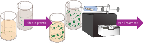
Figure 1. Workflow of media optimization
Results
The phenotypes of the three investigated strains varied significantly depending on the growth medium. All strains demonstrated substantial metabolic activity during both the six-hour pre-growth phase and the post-treatment period. In both LB semi-solid agar and SCFM, tobramycin treatments resulted in a concentration-dependent reduction of metabolic activity and delay in peaking, consistent with typical biofilm behavior (Figs. 2 and 3). Notably, the clinical CF isolates exhibited a decreased response to tobramycin when grown in SCFM compared with LB-agar. This effect was not observed in laboratory strains such as PA14 (Fig. 3). These findings demonstrate that SCFM profoundly influences antimicrobial efficacy and that the calScreener can successfully detect the kinetic effects of treatments on biofilm aggregates in situ within a clinically relevant environment.
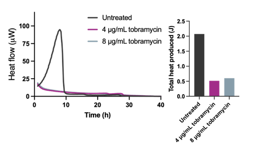
Figure 2. Biofilm aggregate grown in LB-based agar could be completely eradicated by both 4 and 8 µg/mL tobramycin as the clinical strain did not exhibit tolerant pathogenesis in the clinically irrelevant media. The calScreener detected the decline in metabolic activity of all the cells in the aggregates as the antibiotic killed the cells.
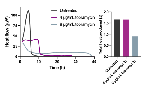
Figure 3. Subpopulations in biofilm aggregates grown in SCFM survived both 4 and 8 mg/mL tobramycin. Although 4 µg/mL did affect the aggregated population, the total metabolism was not altered from the untreated sample – it merely lowered the population output. The calScreener detected the surviving metabolic slow subpopulation in aggregates treated with 8 mg/mL, seen as a prolonged, low metabolic signal.
Conclusions
The Symcel calScreener, used in combination with synthetic CF Sputum Media, represents a powerful tool for advancing the study of cystic fibrosis-related biofilm infections. By recreating complex conditions characteristic of CF infections, the system enables real-time kinetic monitoring and in situ analysis without disrupting biofilm structure.
This method effectively captures clinically relevant treatment responses on biofilms in SCFM, highlighting the importance of using physiologically relevant media. For instance, biofilms grown in SCFM show reduced response to tobramycin compared to those grown in LB agar, underscoring how conventional laboratory media may produce misleading conclusions regarding antimicrobial efficacy. By offering a more realistic model of CF infections, the calScreener has the potential to improve the translational value of biofilm research, optimize resource use, and increase the likelihood of successful clinical interventions.
The need for physiologically relevant, in vivo-like assays is not limited to CF research. With the use of microcalorimetry, models are not limited by their complexity but rather by the imagination of the researchers. The importance of bridging the gap between in vitro and in vivo will not decrease.
REFERENCES
1. Kragh KN, Tolker-Nielsen T, Lichtenberg M. (2023) The non-attached biofilm aggregate. Commun Biol. DOI: 10.1038/s42003-023-05281-4
2. Lewin GR, Kapur A, Cornforth DM, et al. Application of a quantitative framework to improve the accuracy of a bacterial infection model. Proc Natl Acad Sci USA. DOI:10.1073/pnas.2221542120
3. Rumbaugh KP, Bjarnsholt T. Microbial Primer: In vivo biofilm. Microbiology DOI:10.1099/mic.0.001407
FEATURED PRODUCTS